|
The
National Human Neural Stem Cell Resource provides neural
stem cells harvested from the post-natal, post-mortem,
human and animal brain to the research community for stem cell research.
Several brain areas as well as cultures from normal and
genetically mutant specimens are represented in the Resource. Cells
derived from other organ systems are also available.
Stem
cell research will increase our understanding of the nervous
system and may allow us to develop treatments for currently
incurable brain diseases and injuries. The
Resource encourages stem cell research for the study of
these neural stem cells as potential transplantable tissue
for the repair of injury such as that sustained during
traumatic brain injury or stroke, for the repair of pathological
processes such as those seen in the neurogenetic diseases
Hurler's disease or Leigh's disease, or for repair
of neurodegenerative processes such as those seen in Parkinson's
or Alzheimer Diseases.
In addition, the stem cells should be used for stem cell
research aimed at the detailed study of mechanisms of neural
differentiation and transdifferentiation and the genetic
and environmental signals that direct the specialization
of the cells into particular cell types.
Philip H. Schwartz, Ph.D., Director
[email protected] |
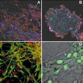
A)
A primary culture of proliferating cells showed a high proportion
of nestin (red) to GFAP (blue) staining in monolayer culture;
co-staining was relatively frequent (10x).
B) Budding neurosphere showed nestin cells (red) at the
circumference with a much higher proportion of GFAP (blue)
to nestin in the interior of the sphere (10x).
C) Nestin (green) and Sox2 (red) staining in proliferating
hNPCs (40x). Arrows show occasional nestin-negative/Sox2-positive
cells.
D) Doublecortin (DCX) staining of hNPCs revealed a subpopulation
of small, 5 - 10 um diameter, DCX-positive (green) cells
against a phase-contrast background (40x).
|